In the first time-resolved 3D study, researchers have examined microstructural changes to dentine caused by acid and found that acid dissolves the minerals in different dentine structures at different rates. (Image: thitinonjong/Shutterstock)
GUILDFORD, UK: Researchers from the universities of Surrey and Birmingham in the UK have recently examined microstructural changes to dentinal tubules during acid demineralisation. The findings aim to provide new insights into conditions such as dental erosion and caries and to help develop new treatments that can restore the structure and function of dentine.
In the study, the researchers employed in situ synchrotron X-ray micro-tomography, which was performed at Diamond Light Source, the UK’s national synchrotron science facility. The electrons were accelerated close to the speed of light to generate X-rays that were used to scan dentine samples during their exposure to acid over 6 hours. In this manner, the researchers were able to create 3D images of dentine’s internal structure with sub-micrometre resolution for the duration of the experiment. From their analysis, they found that acid dissolves the minerals in different dentine structures at different rates.
“At the micro-scale, dentine has tubules that run to the centre of the tooth, and the material lining the tubules, called peritubular dentine, is much more mineralised than the material between the tubules, called intertubular dentine. It was known that peritubular dentine demineralises at a faster rate than intertubular dentine. However, owing to the limitations of laboratory techniques, it was only possible to measure the demineralisation rates and the microstructural changes during the first few minutes of demineralisation and to limited depths,” co-author Nathanael Leung, a final year PhD student at the University of Surrey, told Dental Tribune International.
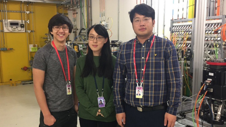
Members of the research team at Diamond Light Source (from left): Nathanael Leung, Dr Tan Sui and Bin Zhu. (Image: University of Surrey)
“With synchrotron X-rays, we were able to measure the demineralisation rates and the microstructural changes of peritubular and intertubular dentine in 4D with high resolution, in a much larger volume of dentine and over a longer time frame,” he continued.
Commenting on the findings, senior author Dr Tan Sui, a senior lecturer in materials engineering at the University of Surrey, said in a press release: “Relatively little is known about how exactly acid damages the dentine inside our teeth at a microstructural level. This new research technique changes that and opens the possibility of helping identify new ways to protect dental tissues and develop new treatments.”
Leung has been awarded a 2021 GSK Award by the Oral and Dental Research Trust and will continue to further investigate the topic. “With the ability to measure the microstructure of dentine, it is possible to create high-fidelity computational models of dentine that can be used to simulate its mechanical response to masticatory forces in correlation to the microstructural changes caused by acid demineralisation. Obtaining the capabilities to faithfully simulate the mechanical response of dentine to different treatments, such as fillings and crowns, might make it possible to improve the methods of these treatments, which can potentially lead to more successful outcomes,” he concluded.
Our teeth are constantly exposed to mechanical and chemical wear, and with age, our oral immune system becomes less effective at fighting pathogenic ...
Education
Live webinar Mon. 13 July 2026 4:30 pm UTC (London)
BIRMINGHAM, England: Social media is increasingly used to promote orthodontic treatment directly to prospective patients, including by direct-to-consumer ...
One year after I argued that dental practices that adopt digital technologies early will dominate tomorrow, artificial intelligence (AI) in dentistry has ...
LONDON, England: Although dietary interventions have been associated with reduced systemic inflammation, their relevance to periodontal disease remains ...

 International / International
 Brazil / Brasil
 Canada / Canada
 Latin America / Latinoamérica
 USA / USA
 Austria / Österreich
 Bosnia and Herzegovina / Босна и Херцеговина
 Bulgaria / България
 Croatia / Hrvatska
 Czech Republic & Slovakia / Česká republika & Slovensko
 France / France
 Germany / Deutschland
 Greece / ΕΛΛΑΔΑ
 Hungary / Hungary
 Italy / Italia
 Netherlands / Nederland
 Nordic / Nordic
 Poland / Polska
 Portugal / Portugal
 Romania & Moldova / România & Moldova
 Slovenia / Slovenija
 Serbia & Montenegro / Србија и Црна Гора
 Spain / España
 Switzerland / Schweiz
 Turkey / Türkiye
 China / 中国
 India / भारत गणराज्य
 Pakistan / Pākistān
 Vietnam / Việt Nam
 ASEAN / ASEAN
 Israel / מְדִינַת יִשְׂרָאֵל
 Algeria, Morocco & Tunisia / الجزائر والمغرب وتونس
 Middle East / Middle East

 Dr. Fernando FranchLive webinar
 Dr. Nicolas OuelletRegister now1CELive webinar

 Dr. Nisha D’Silva BDS, MSD, PhD, Dr. Kıvanç Bektaş-KayhanRegister now1CELive webinar
 Federico ZunicaRegister now1CE


To post a reply please login or register