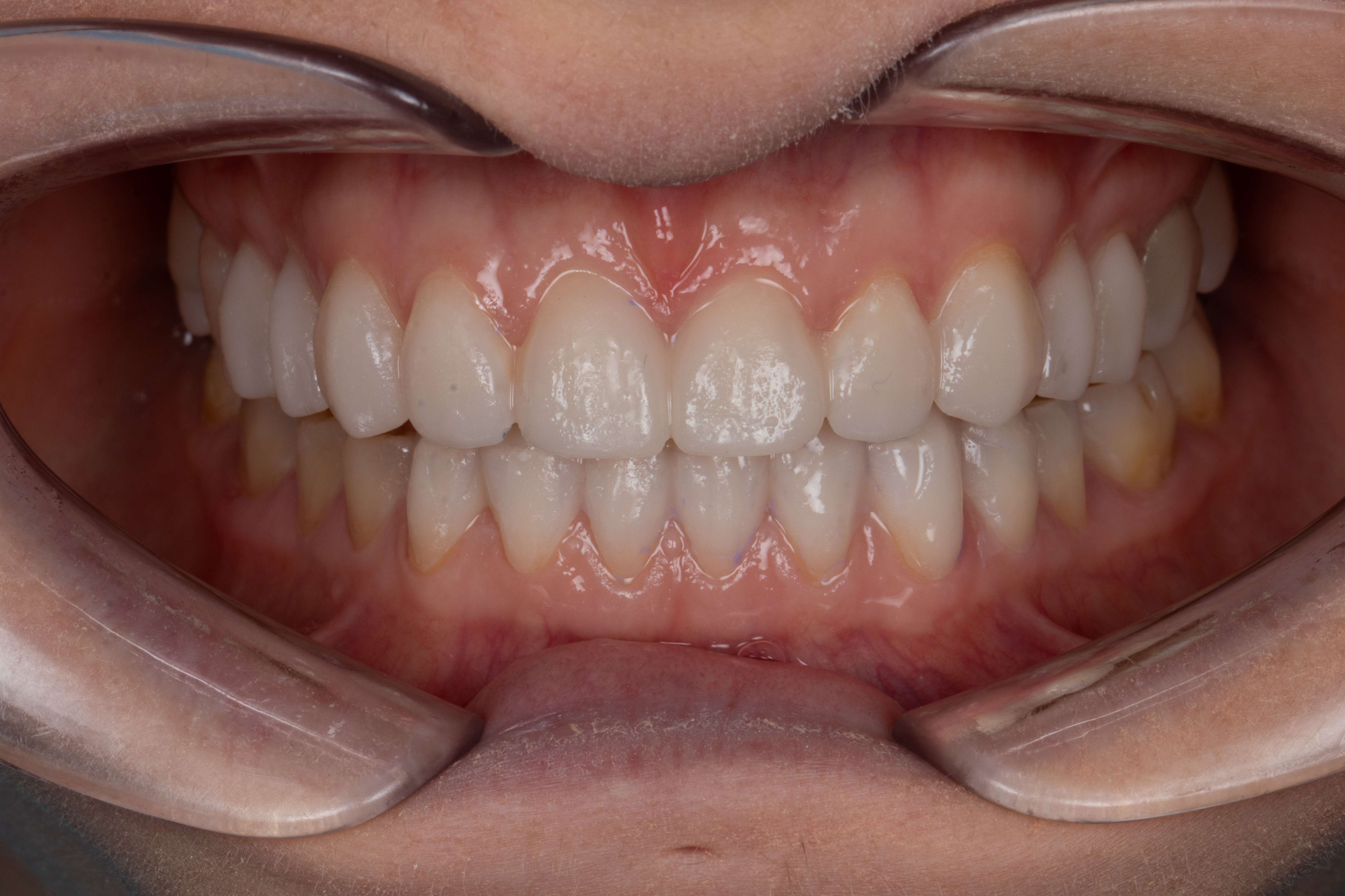
Fig. 1: Extra-oral frontal view before treatment showing shortened and flattened maxillary anterior crowns and their effect on the patient’s smile aesthetics. (All images: Dr Zohaib Ali)
Erosive tooth wear is common in the general population, although global prevalence estimates remain difficult to establish owing to differences in diagnostic criteria and study methodologies.1 Erosive tooth wear is a multifactorial condition that results from the chemical softening of dental hard tissues by intrinsic or extrinsic acids and can significantly accelerate structural loss when combined with mechanical forces such as attrition and abrasion.2–4 Parafunctional habits, most notably bruxism, can further compound this process, leading to complex patterns of generalised wear that affect both function and aesthetics.
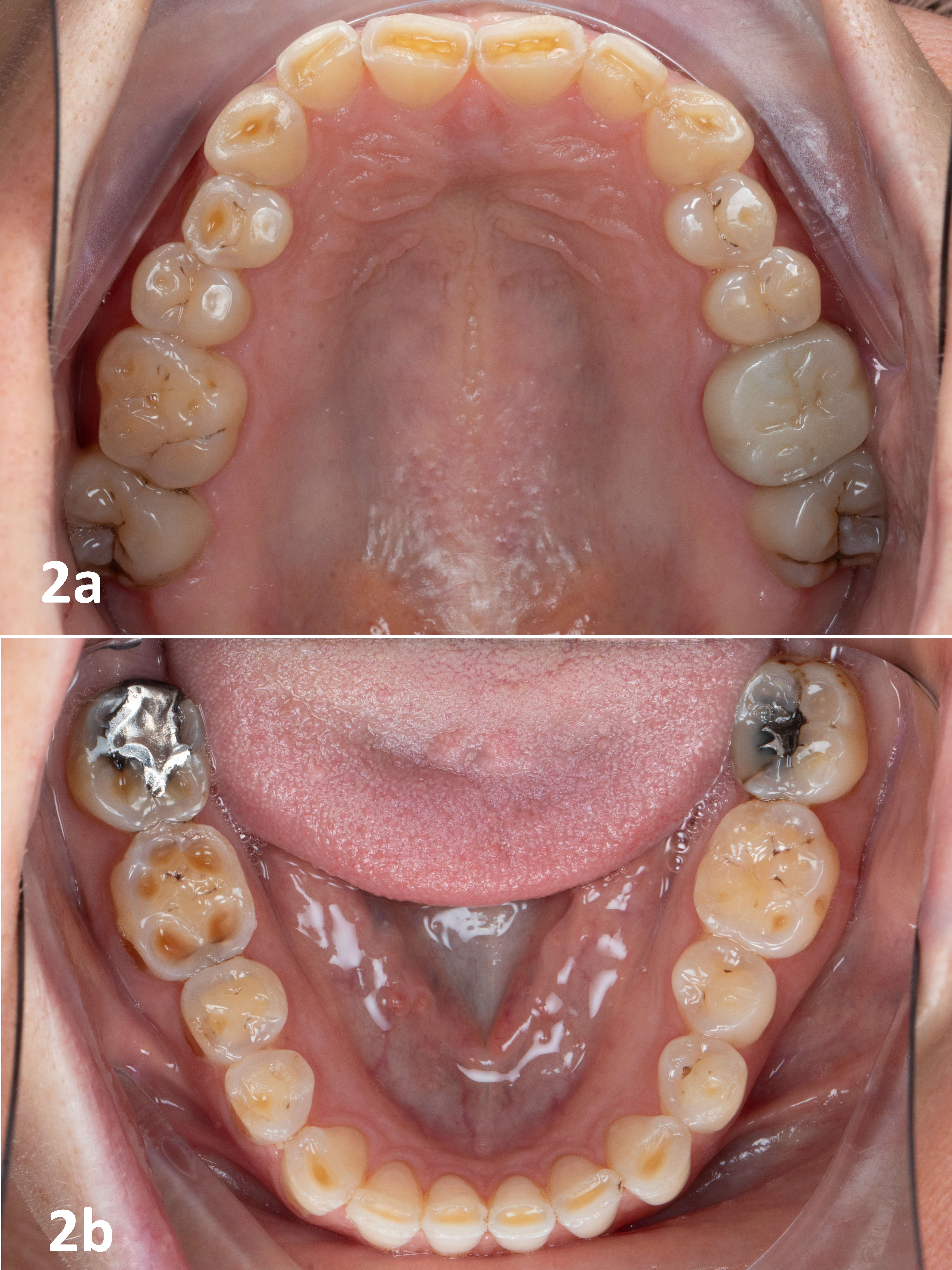
Figs. 2a & b: Intra-oral occlusal views before treatment showing incisal grooving and pronounced occlusal cupping, consistent with an erosive component of the tooth wear. The right side was more severely affected than the left, possibly indicating asymmetric occlusal loading.
Effective management requires a careful balance between restoring occlusal stability, improving aesthetics and preserving the remaining tooth structure. Minimally invasive, adhesive-based strategies have therefore become central to contemporary treatment planning. This case report demonstrates a conservative, adhesive-based rehabilitation approach that integrates direct and indirect adhesive restorations to achieve functional, durable and aesthetic outcomes while respecting the principle of maximal tissue preservation.
Clinical presentation
A young adult woman presented to the dental practice with functional impairment and aesthetic concerns owing to advanced tooth wear (Fig. 1). Her history revealed high dietary acid exposure and both daytime and nocturnal bruxism. Clinical examination showed severe generalised tooth wear with clear erosive and attritional components (Figs. 2a & b, 3a–c). The combination of erosive softening and attrition had exacerbated the tooth wear, resulting in short clinical crowns and a flattened smile curvature. The dentition showed extensive erosive substance loss, characterised by pronounced occlusal cupping and distinct incisal grooving. Several restorations appeared raised owing to the progressive loss of surrounding tooth structure, and broad, flattened buccal enamel lesions further indicated advanced wear.
Notably, the amalgam restorations appeared shiny and untarnished. Other findings unrelated to tooth wear included multiple failing restorations, among them a full-coverage crown on tooth #26 that required replacement. Overall, the acidic diet and parafunctional activity constituted significant contributing risk factors.
Figs. 3a–c: Intra-oral buccal views before treatment showing broad, flattened areas of tooth wear on the buccal surfaces.
Fig. 3b
Fig. 3c
Treatment objectives and rationale
The primary treatment objectives were to restore function and aesthetics while protecting the remaining tooth structure through a minimally invasive approach. A controlled increase in occlusal vertical dimension (OVD) was planned and carefully tailored to the patient’s tolerance to ensure comfort and functional stability. Maintainability and reparability were also key considerations and guided the choice of materials and restorative strategies.
To achieve these goals, a predominantly additive treatment plan was selected to preserve the residual enamel and dentine. Direct composite resin was prioritised owing to its conservative nature and ease of repair and was complemented by indirect restorations in high-load posterior regions. A single zirconia crown was selected to restore the structurally compromised molar, allowing for a conservative vertical preparation.5 This material choice supported a minimally invasive approach and provided the strength and long-term durability required for posterior function.
Fig. 4: Intra-oral mock-up used in the facially driven workflow to assess the planned length of the anterior teeth.
Treatment overview
The full-mouth rehabilitation was carried out at an increased OVD using a predominantly additive approach. The length of the anterior teeth was tested using an intra-oral mock-up (Fig. 4). Treatment began with the maxillary and mandibular anterior teeth, which were restored on the same day. To maintain the newly established vertical dimension during this phase, posterior occlusion was stabilised using temporary composite occlusal stops.
A combined protocol using G-ænial A’CHORD and G-ænial Universal Injectable (both GC) was employed for the anterior restorations. A bilaminar technique was followed. A palatal stent was used to create an initial shell, followed by a dentine body layer using G-ænial A’CHORD and a final enamel layer applied through injection moulding using G-ænial Universal Injectable (Figs. 5a–c).
Four days later, the maxillary premolars and molars were restored using an injection-moulding technique with G-ænial Universal Injectable. The mandibular premolars were restored ten days later. The lower left quadrant was rehabilitated with semi-indirect composite onlays fabricated by injection moulding using G-ænial Universal Injectable (Figs. 6a & b), providing a balance of tooth preservation, cost-effectiveness and long-term repairability.
Ten days later, the maxillary left first molar was restored with a zirconia crown after vertical preparation, providing durable reinforcement for this strategically important posterior tooth. Finally, lithium disilicate onlays (Initial LiSi Press, GC) were placed on tooth #46 and tooth #47 to provide enhanced strength and predictable adhesive performance in an area subject to high occlusal loading. These restorations were completed three months later owing to patient-related scheduling constraints.
Figs. 5a–c: Bilaminar technique used for the anterior restorations under rubber dam isolation. Initial shell and dentine body layer created using G-ænial A’CHORD (a & b). Completed anterior restorations after application of the final enamel layer using G-ænial Universal Injectable (c).
Fig. 5b
Fig. 6a: Semi-indirect composite restorations fabricated by injection moulding using G-ænial Universal Injectable. Restorations on the printed model.
Fig. 6b: Isolation of the teeth before luting.
Adhesive and isolation protocol
All direct composite procedures were performed under rubber dam isolation to ensure optimal control of the operative field and maximise adhesive reliability. A combined layering strategy was employed. Conventional paste composite was used for dentine and enamel replacement, and injection-moulding techniques were incorporated to refine the final contour, improve precision and streamline the workflow.
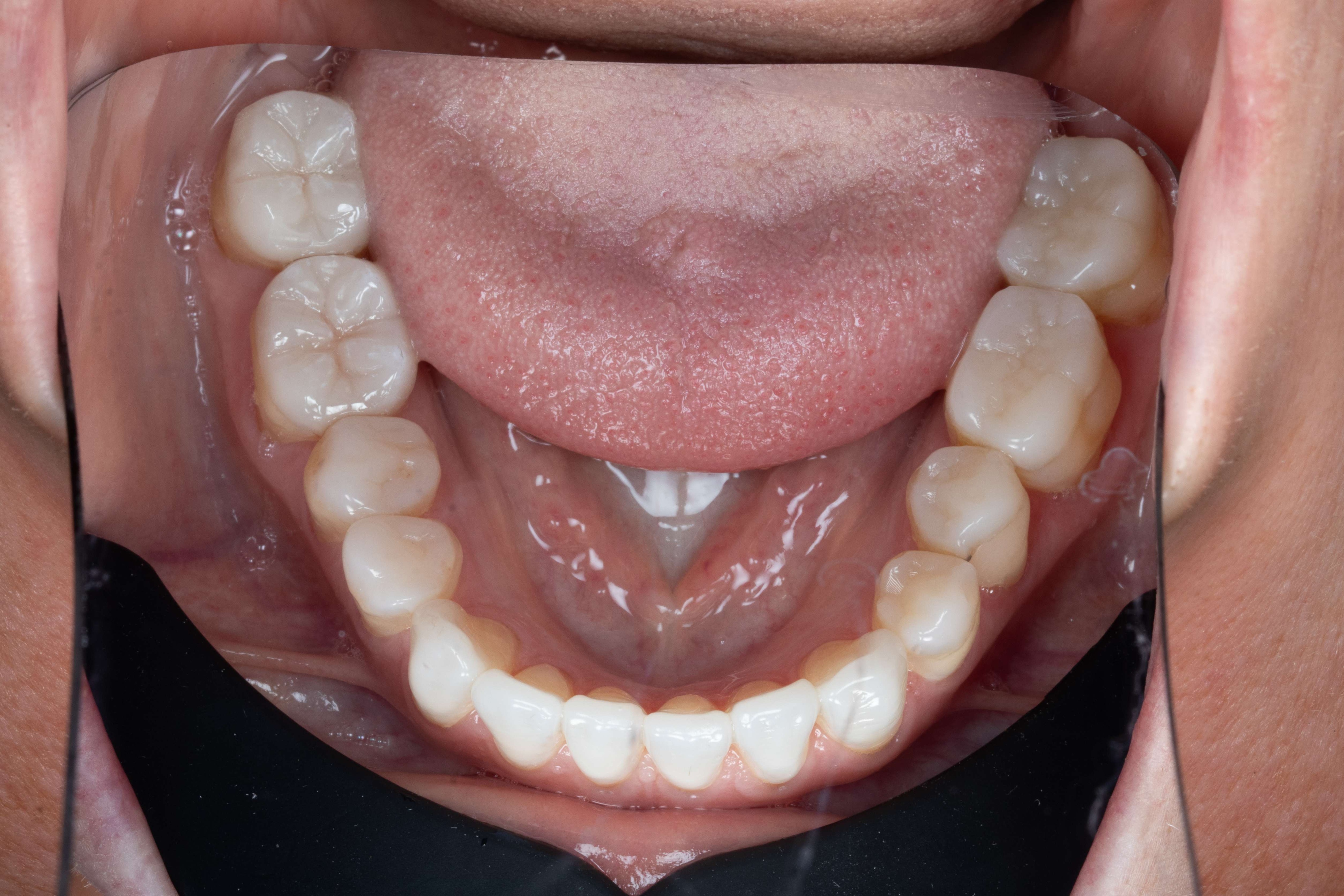
Fig. 7: Occlusal view of the mandibular arch after treatment, showing the polished Initial LiSi Press restorations on tooth #46 and tooth #47.
Adhesive protocols were followed according to the manufacturers’ instructions, and substrate-specific conditioning and bonding procedures were applied to ensure durable interfaces across both direct and indirect restorations. Direct restorations were bonded using G-Premio BOND (GC).
Indirect composite restorations fabricated using G-ænial Universal Injectable were gently sandblasted with 27 μm aluminium oxide and cleaned with alcohol before being silanised using G-Multi PRIMER (GC). The Initial LiSi Press restorations were cleaned with alcohol, etched with hydrofluoric acid for 20 seconds and subsequently silanised using G-Multi PRIMER. Prepared tooth surfaces were cleaned and lightly air-abraded, followed by selective phosphoric acid etching and application of G-CEM ONE Adhesive Enhancing Primer. Final cementation of all glass-ceramic and composite restorations was performed using G-CEM ONE (GC). The zirconia crown was likewise cleaned and lightly sandblasted before cementation using G-CEM ONE.
Occlusion and vertical dimension
The OVD was increased in a controlled manner, allowing ongoing assessment of the patient’s phonetics, comfort and functional adaptation. The new OVD was established through a facially driven workflow that defined the ideal incisal edge position. Subsequent refinements were guided by the patient’s functional response.
Given the available interocclusal space and the patient’s bruxism, a more conservative occlusal anatomy was adopted to reduce the risk of interferences and chipping. In the posterior region, the priority was durable function rather than aesthetics. The lithium disilicate restorations on tooth #46 and tooth #47 were therefore meticulously polished and left unglazed to minimise antagonist wear6,7 (Fig. 7).
Figs. 8a–c: Intra-oral buccal views after treatment showing the restored anterior and posterior dentition and the established occlusal relationship.
Fig. 8b
Fig. 8c
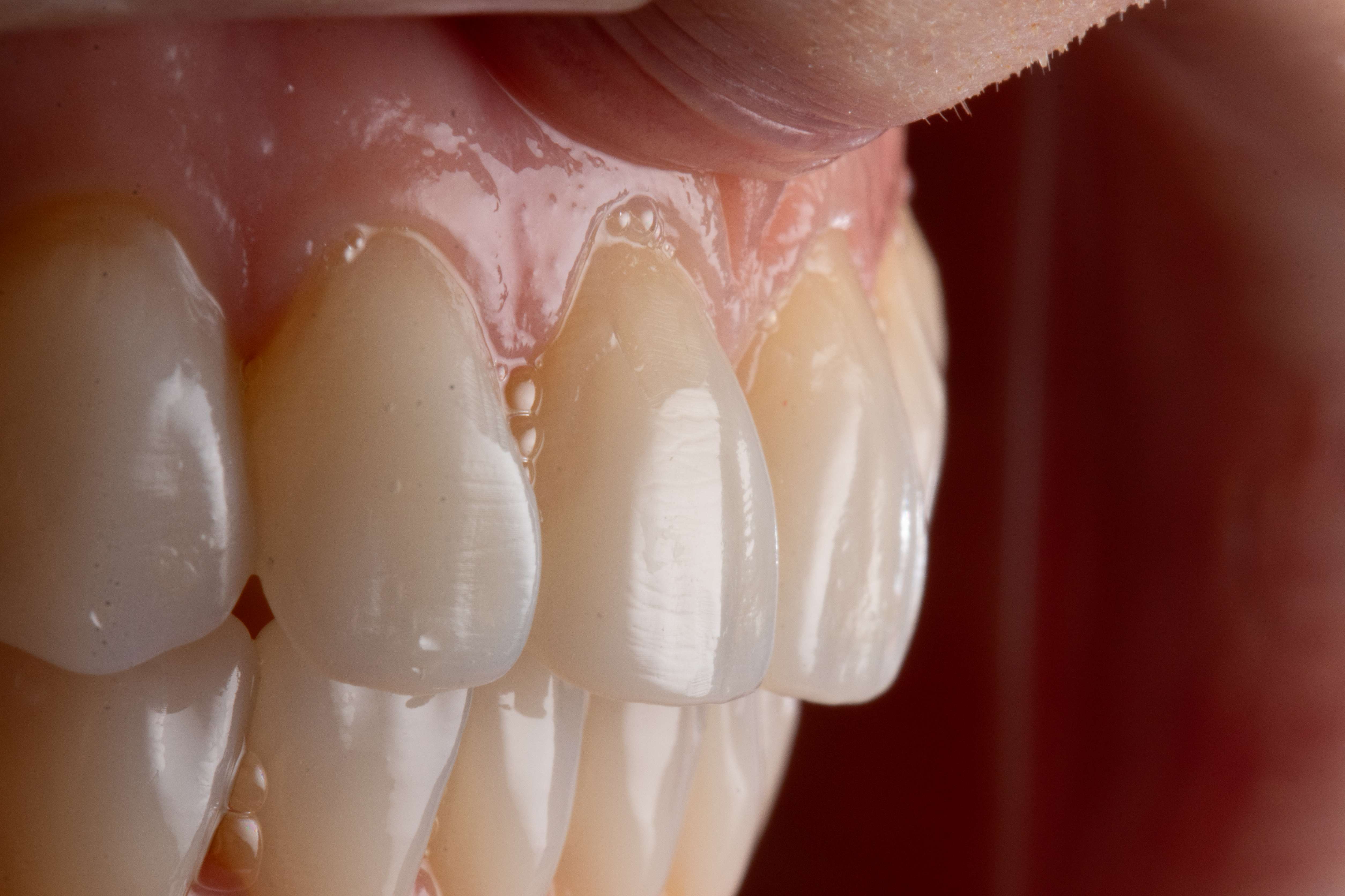
Fig. 9: Close-up view of the maxillary anterior restorations showing the restored surface morphology and texture.
The final occlusal scheme provided stable bilateral posterior contacts and anterior guidance that limited posterior interferences during excursive movements. This configuration respected the patient’s functional envelope and promoted a predictable and comfortable occlusal relationship.
Outcomes
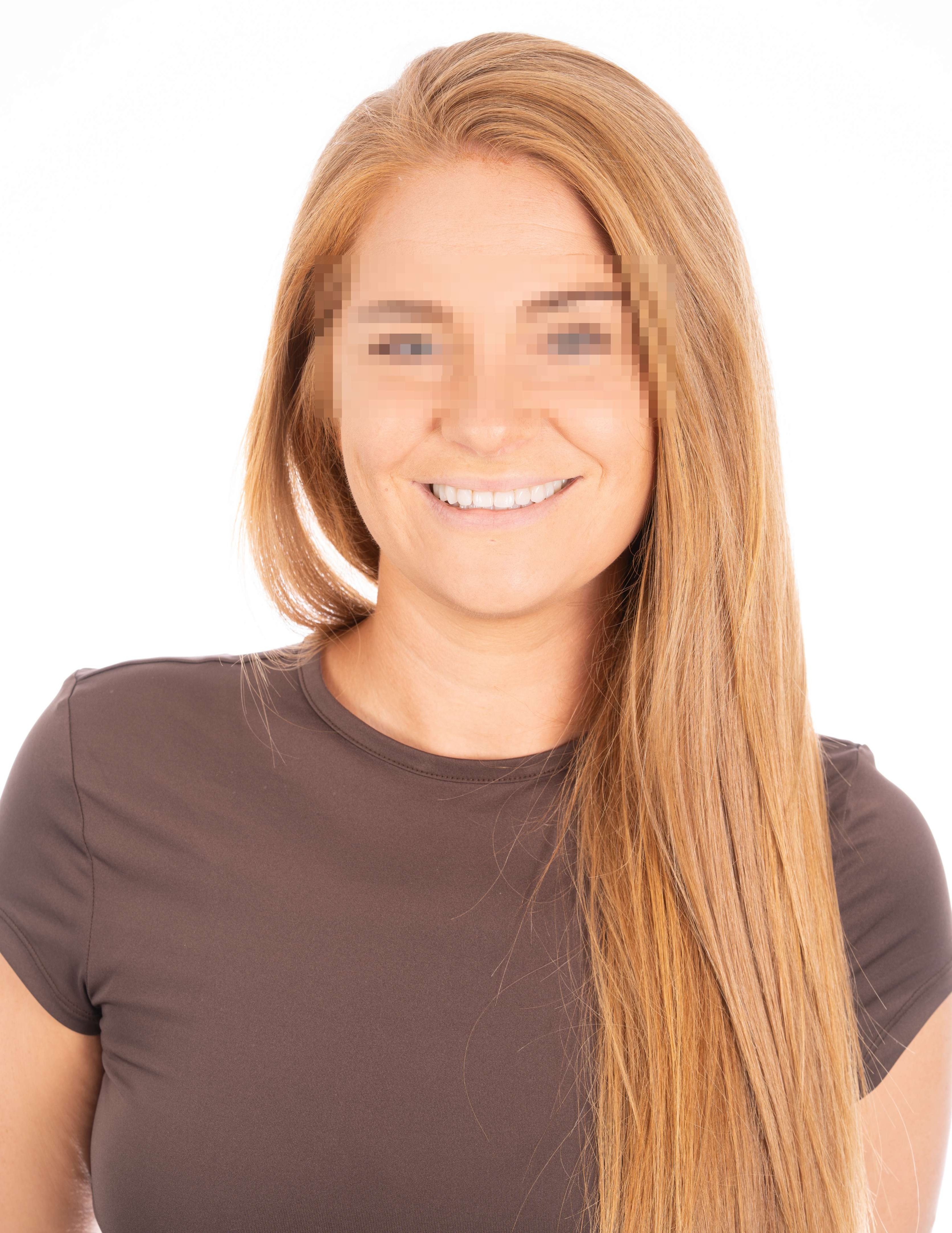
After completion of treatment, the patient demonstrated stable occlusion at the newly established OVD (Figs. 8a–c). The additive, minimally invasive approach preserved existing tooth structure while providing comprehensive coverage of the worn surfaces. Aesthetic outcomes were enhanced through harmonised anterior composite layering (Figs. 9 & 10), and posterior morphology was restored using direct, indirect and semi-indirect restorative modalities. The predominance of composite in the rehabilitative strategy supports long-term maintainability because composite materials allow straightforward polishing and repair as well as staged restorative refinement if required.
Maintenance and risk management
Fig. 10: Extra-oral frontal view after treatment showing the restored anterior tooth length and smile curvature, with the maxillary incisal edges following the curvature of the lower lip.
The long-term success of the rehabilitation was supported by a tailored maintenance programme. The patient received counselling on dietary modifications aimed at reducing erosive challenges as well as behavioural guidance to address parafunctional activity associated with bruxism. Protective therapy included ongoing use and periodic adjustment of an occlusal appliance to mitigate excessive loading on the restorations. Regular professional reviews were scheduled to provide hygiene support, monitor the patient’s adaptation to the increased OVD and maintain the integrity of the adhesive interfaces, including polishing or repairing composite surfaces as needed.
Discussion
This case demonstrates that conservative, adhesive-based full-mouth rehabilitation can effectively address severe erosive tooth wear while preserving tooth structure. Materials were carefully selected based on their respective indications. Most of the rehabilitation was completed using composite resin, selected for its minimally invasive nature, cost-effectiveness and repairability, making it particularly suitable for long-term maintenance in a young patient.
A full-coverage zirconia crown was placed on tooth #26 to allow minimally invasive vertical preparation and provide the strength and durability required to restore this structurally compromised molar. In contrast, the partial restorations in the lower right quadrant were fabricated from lithium disilicate, selected for its reliable adhesive bonding performance and favourable strength profile and providing a balance between conservation of tooth structure and functional longevity.
Molar incisor hypomineralisation (MIH), recognised as a clinical condition since 2001, is a common developmental enamel defect affecting the first permanent...
LEUVEN, Belgium: Whether it is a sophisticated composite or an advanced adhesive, every new dental product must be safe, precise and reliable. At GC, ...
LEUVEN, Belgium: GC Corporation is marking a special anniversary in the dental world this year as it looks back on 105 years of history. What began in 1921 ...
LEUVEN, Belgium: GC Europe, a leading oral health company supplying dental products and services to dental professionals, is emphasising the importance of ...
Education
Live webinar Thu. 5 November 2026 6:00 pm UTC (London)
TOKYO, Japan: Leading dental manufacturer GC Corp. has invited global dental professionals to save the date for its sixth International Dental Symposium, ...
As restorative dentistry shifts focus from core operative procedures to individual patient needs and values, the demand for holistic patient care for ...



 International / International
 Brazil / Brasil
 Canada / Canada
 Latin America / Latinoamérica
 USA / USA
 Austria / Österreich
 Bosnia and Herzegovina / Босна и Херцеговина
 Bulgaria / България
 Croatia / Hrvatska
 Czech Republic & Slovakia / Česká republika & Slovensko
 France / France
 Germany / Deutschland
 Greece / ΕΛΛΑΔΑ
 Hungary / Hungary
 Italy / Italia
 Netherlands / Nederland
 Nordic / Nordic
 Poland / Polska
 Portugal / Portugal
 Romania & Moldova / România & Moldova
 Slovenia / Slovenija
 Serbia & Montenegro / Србија и Црна Гора
 Spain / España
 Switzerland / Schweiz
 Turkey / Türkiye
 China / 中国
 India / भारत गणराज्य
 Pakistan / Pākistān
 Vietnam / Việt Nam
 ASEAN / ASEAN
 Israel / מְדִינַת יִשְׂרָאֵל
 Algeria, Morocco & Tunisia / الجزائر والمغرب وتونس
 Middle East / Middle East

 Dr. Dimple DesaiRegister now1CELive webinar
 Dr. Ahmed ElmoghazyLive webinar
 Dr. Gil Asafrana D.M.D, B.M.ScRegister now1CELive webinar
 Dr. Adrian Gnatek PhD | Oral SurgeonRegister now1CE























To post a reply please login or register